


What is an X-ray machine, and what are the applications of X-rays in medicine?
X-ray machines use X-ray beams to create images of the internal structures of the human body. These images help doctors and medical staff examine the structure of organs, bones, blood vessels, and other parts of the body to diagnose and monitor health problems such as fractures, lung infections, cancer, etc.
1. A brief history of its formation.
The first person The first person to discover X-rays in medicine was Wilhelm Röntgen (often called Rơnghen in Vietnam), a German physicist. He discovered X-rays on November 8, 1895. After leaving the laboratory, he suddenly remembered he had forgotten to switch off the high-voltage power supply to the cathode ray tube. Wilhelm Conrad Röntgen returned to the room and suddenly saw a greenish light on the table, even though the room was pitch black. Noticing this strange phenomenon, he stayed in the laboratory for 49 days and eventually discovered the properties of this mysterious ray, which he tentatively named X-rays.
Wilhelm Röntgen He conducted experiments with X-rays and used them to create the first images of bones and objects within the human body. This opened the door to modern medicine and diagnostic imaging and made a significant contribution to medicine and other related fields. Therefore, Wilhelm Röntgen is recognized as a pioneer in the application of X-rays in medicine with the X-ray machine.
The first X-ray machine X-rays are used worldwide, originating in Germany. After Wilhelm Röntgen discovered X-rays in 1895, he quickly conducted experiments with this beam and created the first images of bones and objects within the human body.
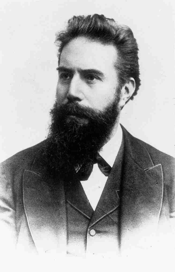
Röntgen A pioneer in the application of X-rays with X-ray machines in medicine.
2. Applications of X-rays in medical X-ray imaging:
2.1 Diagnostic imagingX-ray machines use X-ray beams to create images of the internal structures of the human body. These images help doctors and medical staff examine the structure of internal organs, bones, blood vessels, and other body parts to diagnose and monitor health problems such as fractures, lung infections, cancer, etc.
2.2 Magnetic Resonance Imaging (CT) ScanA CT scan, also known as a CAT scan, uses X-ray beams and special sensors to create images that pass through thin slices of the body. This allows for viewing 3D structures and detecting internal body problems in greater detail.
2.3 Supportive treatmentX-ray beams can be used to assist in guiding medical interventions, such as performing procedures like aspiration and injection protocols in real time with the aid of X-ray imaging.
2.4 Mobile X-ray: Mobile X-ray devices are used in emergency situations or when X-rays are needed right at the patient's bedside, saving time and providing convenience for patients.
3 – Structure of an X-ray machine:
In terms of structure, X-ray machines differ considerably between generations, but basically consist of the following parts:
– X-ray generator.
– High voltage generator.
– Control unit.
– Image acquisition/display unit.
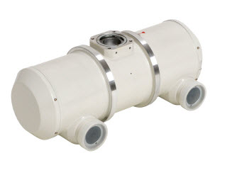
X-ray tube

X-ray image receiver
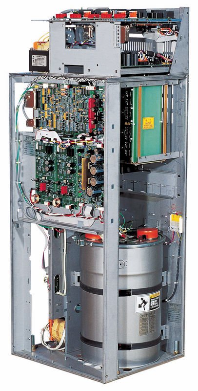
High Voltage Unit
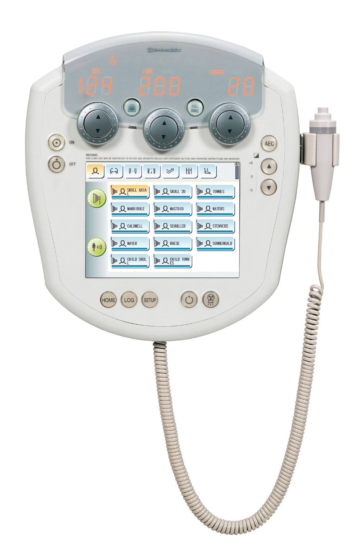
X-ray Machine Control Panel
4 – Classification of X-ray machines
X-ray machines can be classified based on various criteria, including purpose, structure, operation, and specific applications in medicine or industry. Below are some of the main classifications of X-ray machines.
4.1 According to image processing technology:
– Traditional X-ray (using film), projected X-ray (screen), indirect digital X-ray (CR), direct digital X-ray (DR).
Nowadays, traditional X-ray machines using negative film are rarely used due to several factors: radiation safety issues, environmental hygiene, inconvenience, etc. Instead, digital X-ray machines are gradually replacing them due to many advantages: safer, images are captured in digital form, saved to a computer, and easily edited.
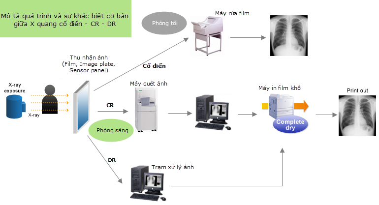
Classical X-ray: X-ray imaging (X-ray film/intensifying film) is used to image body parts. The film is stored in a cassette. The cassette is placed behind the object to be imaged; after passing through the object, the X-rays strike the film. After exposure, the film is taken to a darkroom for processing with developing chemicals. When developing the film, AgCl is used. Areas that reacted with X-rays will not be lost during development (resulting in black), while areas that did not react (such as bone, where X-rays are blocked) will be lost during development (resulting in white). The image is then read on a film reader. This is a permanent image, which cannot be altered, making it difficult to store, copy, and retrieve.
Indirect digital X-ray (CR) Computed Radiography (CRI) is a system similar to traditional X-ray, but instead of a standard X-ray machine and film/intensifying plate, it uses an imaging plate coated with a phosphor layer that stores and stimulates luminescence. When X-rays are shone onto the imaging plate, a latent image is created. This image is then scanned a second time by a laser in a digitizer, capturing the light and producing a digital image (a conversion from analog to digital). This image is then processed by a computer. The imaging plate is erased by a white light source and reused. The number of reuses depends on the technology, materials, and manufacturer of the imaging plate.
Direct Radiography (DR) X-ray:
This technique is similar to digital cameras, as it also uses the same principle of a sensor panel and produces images immediately after shooting. The image creation principle relies on a sensor panel constructed from a combination of a scintillating layer (consisting of cesium iodide/thallium layers) and a transistor film (TFT) with amorphous silicon. This sensor panel replaces the classic film/intensifying plate pair; after exposure, it transfers the image and displays it on a computer screen after about 5 seconds, and can be used for further shooting without erasing like CR.
4.2 According to intended use:
– Medical X-ray machine: Used in the medical field to image the human body and diagnose diseases. It captures detailed images of body parts such as bones, lungs, heart, etc.
– Industrial X-ray machines: Used in industry for product quality inspection and control, welding inspection, defect detection, etc.
Classification based on specific applications in healthcare:
– Magnetic Resonance Imaging (CT Scan): Uses X-rays to create 3D images of organs and internal structures in the body.
– Functional diagnostic imaging machine (Fluoroscopy): Creates continuous moving images of organs and systems in the body, suitable for real-time interventional procedures.
– Tomosynthesis imaging: Creates detailed images of organs and surrounding areas by capturing images from multiple angles and combining them into a 3D image.
The classifications above are just a few examples, and there may be many other criteria for classifying X-ray machines based on specific purposes and usage requirements.
Related articles:
-
Someko Healthineers participate in Mid-Autumn Festival Organizing Activities.
-
24/7 Customer Support – GE Home Surgical Carm System
-
64-Slice Computed Tomography (CT) Scanner System with Integrated AI Technology
-
Someko Healthineers provides remote support for hospitals, including C-arm machines and Cios Connect.
-
AI in Healthcare for Early Diagnosis and Cancer Screening in the 2026 Era.
-
Installation and Handover of CXK MRI 0.35T Magnetic Resonance Imaging System
-
Music by Bao Vq
-

Top C-Arm from SOMEKO Healthineers
-

Super Cup “SOMEKO ChampionTour26”
-
GE C-Arm
-
C-Arm Acadis Varic – Someko Healthineers
-
Super Cup Celebrating National Day, September 2nd
-
Instructions for using the Someko C-arm X-ray machine.
-

Medical X-rays: Advantages, Limitations, and When Are They Needed?
-
What is the price of a genuine DR X-ray image receiver plate?


Thank you for the information.